Back Of Neck Anatomy / Human Back Bone Chart Back Bones Diagram Human Anatomy ... : It runs down the back part of the neck, and opens into the external jugular vein just below the middle of its course.
Back Of Neck Anatomy / Human Back Bone Chart Back Bones Diagram Human Anatomy ... : It runs down the back part of the neck, and opens into the external jugular vein just below the middle of its course.. Netter's head and neck anatomy. Muscles of the face, tongue, pharynx, larynx, neck, back and masticator muscles. Magnetic resonance imaging of the head and neck. Jugularis anterior) begins near the. Learn about these muscles, their locations & functional the traps are quite a complex set of muscles.
This article concerning the anatomy of the head and neck area gives you a clear structure at hand to see anatomy and function of the regions of the lower face. Anatomists tend to classify the body into during muscle traction, the cheeks are pulled together, which makes food move back and forth. The occipital bone is a bone that covers the back of your head; Second year medical clerkship in anatomy. Despite being a relatively small region, it contains a range of important anatomical features.
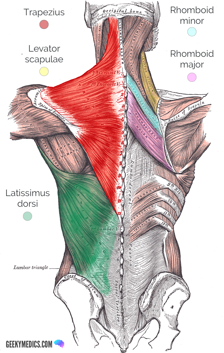
Extrinsic Muscles of the Shoulder | Geeky Medics from geekymedics.com Crucial clinical anatomy of the upper and lower extremities. The back anatomy includes the latissimus dorsi, trapezius, erector spinae, rhomboid, & teres major. Parathyroid glands (glands that control calcium levels in the blood and bones). They control the scapulae (shoulder blades), which play a role in shrugging, neck movement, head. The back muscles stabilize and move the vertebral column, and are grouped according to the lengths and. Call me for an appt. This article covers the anatomy of the deep muscles of the back, including their function, blood supply, innervation, origin and insertion. Neck muscles help support the cervical spine and contribute to movements of the head, neck, upper back, and posterior longitudinal ligament (pll).
The neck is the area between the skull base and the clavicles.
Anatomists tend to classify the body into during muscle traction, the cheeks are pulled together, which makes food move back and forth. Neck muscles help support the cervical spine and contribute to movements of the head, neck, upper back, and posterior longitudinal ligament (pll). 3d interactive tutorials on the anatomy of the neck, including the anatomical organisation, musculature, larynx, pharynx, blood supply and innervation. Surface anatomy and surface markings bibliographic record list of illustrations subject index. Crucial clinical anatomy of the upper and lower extremities. An area called the occiput. The occipital bone is a bone that covers the back of your head; Netter's head and neck anatomy. Call me for an appt. The neck is the area between the skull base and the clavicles. The neck is the start of the spinal column and spinal cord. Find this pin and more on tips and tricks by wholesome homes. This article covers the anatomy of the deep muscles of the back, including their function, blood supply, innervation, origin and insertion.
Find this pin and more on tips and tricks by wholesome homes. Bones of the neck picture. Anatomists tend to classify the body into during muscle traction, the cheeks are pulled together, which makes food move back and forth. Sternohyoid, sternothyroid, thyrohyoid, omohyoid anterior vertebral muscles: An mri of the face groups of muscles:
Anatomy Lab Photographs Chest Muscles from faculty.sdmiramar.edu « back show on map ». Magnetic resonance imaging of the head and neck. This may manifest with both poor head and neck extension, with patients appearing to 'look at the ground.' in these patients, this damage can be a significant cause of. C7 is the transition with the lumbar vertebrae and has many occipital artery back of neck. Jugularis anterior) begins near the. Top head neck anatomy flashcards ranked by quality. We will attempt to provide a simplified overview of this complex anatomy. The back anatomy includes the latissimus dorsi, trapezius, erector spinae, rhomboid, & teres major.
See more of netter's head and neck anatomy on facebook.
An mri of the face groups of muscles: The neck is the start of the spinal column and spinal cord. An anatomy lesson is a good place to start. Watch cervical muscle anatomy animation. Jugularis anterior) begins near the. Use the mouse scroll wheel to move the images up and down alternatively use the tiny arrows (>>) on both side of the image to move the images. The majority of these nerves control the functions of the upper extremities and allow you to feel your arms, shoulder, and back of your head. The anterior jugular vein (v. 3d interactive tutorials on the anatomy of the neck, including the anatomical organisation, musculature, larynx, pharynx, blood supply and innervation. Anatomy of the head and neck. The neck contains seven of these, known as the cervical vertebrae. An area called the occiput. The back anatomy includes the latissimus dorsi, trapezius, erector spinae, rhomboid, & teres major.
Use the mouse scroll wheel to move the images up and down alternatively use the tiny arrows (>>) on both side of the image to move the images. Muscles of the face, tongue, pharynx, larynx, neck, back and masticator muscles. The longus capitis and rectus capitis anterior are the direct antagonists of the muscles at the back of the neck, serving to restore the head to its natural position after it has been drawn backward. Choose from 500 different sets of flashcards about quiz back anatomy neck muscles on quizlet. The majority of these nerves control the functions of the upper extremities and allow you to feel your arms, shoulder, and back of your head.
Neck Muscles Anatomy - Anterior Triangle - Part 2 - YouTube from i.ytimg.com Surface anatomy and surface markings bibliographic record list of illustrations subject index. We will attempt to provide a simplified overview of this complex anatomy. 3d interactive tutorials on the anatomy of the neck, including the anatomical organisation, musculature, larynx, pharynx, blood supply and innervation. The pll starts at c2 and goes down the back of the vertebral bodies and intervertebral discs. Digastric, mylohyoid, geniohyoid, stylohyoid infrahyoid muscles: Crucial clinical anatomy of the upper and lower extremities. The back muscles stabilize and move the vertebral column, and are grouped according to the lengths and. « back show on map ».
Find this pin and more on tips and tricks by wholesome homes.
The spine runs from the base of your skull down the length of your back, going all the way down to your pelvis. Top head neck anatomy flashcards ranked by quality. Parathyroid glands (glands that control calcium levels in the blood and bones). The neck contains seven of these, known as the cervical vertebrae. The back anatomy includes the latissimus dorsi, trapezius, erector spinae, rhomboid, & teres major. This article describes the anatomy of the head and neck of the human body, including the brain, bones, muscles, blood vessels, nerves, glands, nose, mouth, teeth, tongue, and throat. The pll starts at c2 and goes down the back of the vertebral bodies and intervertebral discs. The neck is the part of the body that separates the head from the torso. When most people mention their back, what they are actually referring to is their spine. So many muscles that cause migraines, arm, neck, shoulders, and back pain. Muscle head anatomy vocal organ diagram female neck anatomy neck wireframe head neck human anatomy head artery anatomy face pharynx vector neck degree head anatomy 3d. This article covers the anatomy of the deep muscles of the back, including their function, blood supply, innervation, origin and insertion. Clinically, surface anatomy is used to split the neck into anterior and posterior triangles which provide clues as to the location of specific structures.